During a plenary session at EAN 2019 on the role of inflammation in “non-inflammatory” neurological diseases, one speaker discussed the current evidence for a role of neuroinflammation in the pathogenesis of Parkinson’s disease. Inflammation starts early in the disease process and may change the course of disease. To be successful, trials of anti-inflammatory agents must target early stages of the disease and require long treatment periods to demonstrate any neuroprotective effect.
염증이 비염증성 신경 질환에 미치는 영향을 주제로 한 2019년 유럽신경학회 학술회 총회에서 한 연자는 신경염증이 파킨슨병 발병기전에 미치는 영향에 대한 현행 근거(current evidence)에 대한 논의했습니다. 염증은 병의 진행 초기에 시작되며, 병의 진행에 변화를 줄 수 있습니다. 성공적 치료를 위해서 항염증제 임상연구는 질환 초기를 치료 타깃으로 해야 하며, 신경 보호 효과를 입증하려면 장기적인 치료가 필요합니다.
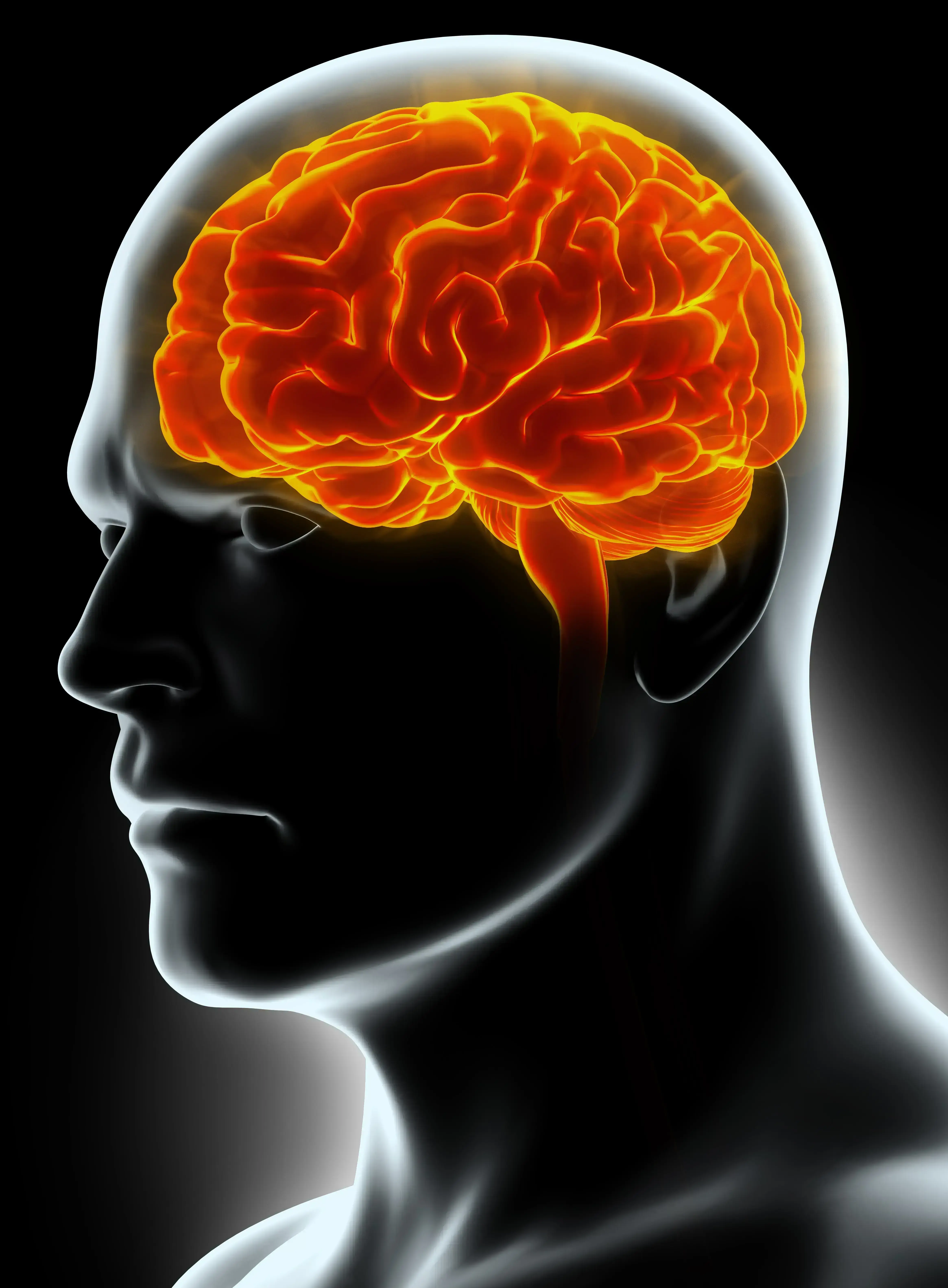
The major pathological features of Parkinson’s disease (PD) – the most common neurodegenerative movement disorder – are the death of substantia nigra dopaminergic neurons and the presence of intraneuronal aggregates (Lewy bodies) composed of α-synuclein.
가장 흔한 신경퇴행성 운동 장애인 파킨슨병의 주요 병리학적 특성은 흑질 도파민 뉴런의 소멸과 알파-시뉴클레인으로 구성된 뉴런 내 집합체(루이소체)의 형성입니다. 가장 흔한 신경퇴행성 운동 장애인 파킨슨병의 주요 병리학적 특성은 흑질 도파민 뉴런의 소멸과 알파-시뉴클레인으로 구성된 뉴런 내 집합체(루이소체)의 형성입니다.
Approximately ∼30% loss of dopamine cell bodies in the substantia nigra and ∼50% loss of dopaminergic axon terminals in the putamen are usually detected at the time of diagnosis of PD suggesting that this level of dopamine reduction is required to produce clinical symptoms. Further degeneration of dopamine neurons is rapid, with almost total loss of dopamine axon terminals in the dorsal putamen detected after 4 years and extensive loss of dopamine cell bodies in the substantia nigra by 5 years.1
파킨슨병 진단 시점에 대략적으로 흑질 도파민 세포체는 ~30%, 조가비핵(putamen) 내 도파민 축삭 말단은 ~50%가 소멸되어 있는 경우가 많습니다. 이는 곧 도파민이 이 정도는 감소해야 임상 증상이 나타남을 의미합니다. 도파민 뉴런의 추가적인 퇴행은 빠르게 진행되어 진단 후 4년 경과 시 등쪽 조가비핵(dorsal putamen)의 도파민 축삭 말단이 거의 대부분 소멸되고, 5년 내에 흑질 도파민 세포체의 소실도 활발히 일어나게 됩니다.1
Consequently, if dopamine neurons are to be rescued, neuroprotective treatments must start as soon as possible.
결국, 도파민 뉴런을 보호하려면 신경 보호 치료를 가급적 빨리 시작해야 합니다.
If dopamine neurons are to be rescued, neuroprotective treatments must start early in the disease process
도파민 뉴런을 보호하려면 병이 진행되는 동안 신경 보호 치료를 최대한 조기에 시작해야 합니다
To validate a neuroprotective approach, we first need to examine the contribution of inflammation in the neurodegenerative processes in PD, and also to identify whether inflammatory processes start early enough such that anti-inflammatory treatments can be expected to have neuroprotective effects.
신경 보호 치료법을 검증하기 위해서는 우선 염증이 파킨슨병의 신경퇴행 과정에 어떤 영향을 미치는지, 그리고 항염증제의 신경보호 효과를 충분히 기대할 수 있을 정도로 염증 과정의 시작이 이른지 여부를 여부를 파악해야 합니다.
How does inflammation contribute to the degeneration of neurons in PD?
염증이 파킨슨병의 뉴런 퇴행에 어떻게 기여하는가?
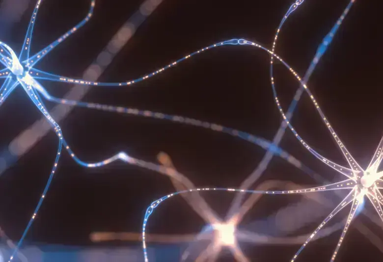
Brain inflammation is characterized by the presence of activated microglia, infiltrating T-lymphocytes, and elevated concentrations of both pro-inflammatory and anti-inflammatory cytokines.
뇌 염증은 활성화된 소교세포, 침윤성 T-림프구의 존재, 그리고 증가된 전염증성 및 항염증성 사이토카인 농도로 특징지어집니다.
Microglia – the “immune cells of the brain” – are known to be activated in the brains of patients with advanced PD. Recent studies show that microglia activation is an early event in PD probably contributing to the development of a-synuclein pathology.2
뇌의 면역세포인 소교세포는 중증 파킨슨병 환자의 뇌에서 활성화되는 것으로 알려져 있습니다. 최근 연구에 따르면 소교세포 활성화는 파킨슨병 환자에게서 조기에 발견되는 현상으로서, 알파-시누클레인 병리 진행에 일조하는 것으로 추정됩니다.2
Microglia activation is an early event in PD probably contributing to the development of a-synuclein pathology
소교세포 활성화는 파킨슨병 초기에 나타나는 현상으로서 알파-시누클레인 병리 진행에 일조하는 것으로 보입니다
Microglia can produce and secrete pro-and anti-inflammatory cytokines, both of which are elevated in the brains of PD patients. However, the overall effect of microglia activation is damaging, with the effect of pro-inflammatory cytokines overshadowing the anti-inflammatory effect and so triggering neurodegeneration.
소교세포는 전염증성 및 항염증성 사이토카인을 생성 및 분비할 수 있으며, 두 종류의 사이토카인 모두 파킨슨병 환자의 뇌에서 증가합니다. 그러나 전염증성 사이토카인의 효과가 항염증성 효과를 압도하여 신경퇴행을 야기하기 때문에 소교세포 활성화의 전반적인 효과는 상해를 일으키는 쪽입니다.
So, what is the trigger of microglia activation and cytokine production? a-synuclein activates microglia through surface receptors and induces a pro-inflammatory response with cytokine production. Following an initial neurotoxic insult, a self-perpetuating cycle of neurotoxicity through microglial activation leading to neuronal death is triggered.3
그렇다면 소교세포 활성화와 사이토카인 생성을 초래하는 요인은 무엇일까요? 바로 알파-시누클레인이 표면 수용체를 통해 소교세포를 활성화하고 사이토카인 생성을 수반하는 전염증성 반응을 유도합니다. 초기의 신경독성 공격이 이루어진 후, 소교세포 활성화를 통해 신경독성이 자가반복(self-perpetuating)되면서 신경세포 사멸이 시작됩니다.3
Inflammation matters: Inflammation starts early and may change the course of PD
염증은 중요합니다 - 염증은 초기에 시작되어 파킨슨병 진행 과정을 변화시킬 수 있습니다.
Could PD be an auto-immune disease?
파킨슨병을 자가면역 질환으로 볼 수 있는가?
Recent evidence suggests a role for the acquired immune system in PD pathogenesis, with the identification of T-lymphocytes in the substantia nigra and a-synuclein reactive T-lymphocytes in the blood of PD patients.4 In addition, the number of major histocompatibility complex (MHC) class II-positive microglia in the subtantia nigra and putamen increase with disease progression in PD patients.5 MHC class II genes encode molecules such as α-synuclein that present the α-synuclein antigen to T-cell receptors generating autoreactive T-lymphocytes. T-cells recognize disease-altered self-proteins as foreign antigens, suggesting that PD could be an auto-immune disease.
최근 연구에서는 파킨슨병 환자에게서 흑질 내 T-림프구와 혈중 알파-시누클레인 반응성 T-림프구가 발견됨에 따라 파킨슨병 발병기전에 후천 면역 체계가 미치는 영향이 있음을 시사했습니다.4 또한, 파킨슨병이 진행됨에 따라 흑질 및 조가비핵 내 주요 조직 적합 유전자 복합체(MHC) II형 양성(class II-positive) 소교세포의 수가 증가합니다.5 MHC II형 유전자는 자가반응 T-림프구를 생성하는 T-세포 수용체에 알파-시누클레인 항원을 제시(present)하는 알파-시누클레인과 같은 분자를 부호화(encode)합니다. T-세포는 질환으로 인해 변화된 자가 단백질을 외부 항원으로 인식하며, 이는 곧 파킨슨병을 자가면역 질환으로 볼 수 있음을 의미합니다.
When in the disease process the priming of T-cells happens is still unclear. Microglia could be activated and present α-synuclein to T-cells prior to neuronal death.
파킨슨병 진행 과정 중 T-세포의 촉발 시기에 대해서는 아직 명확하게 밝혀진 바가 없습니다. 소교세포는 뉴런이 소멸되기 전 활성화되어 알파-시누클레인을 T-세포에 제시할 수 있습니다.
Is the gut the trigger of PD?
소화관은 파킨슨병을 촉발시킬 수 있는가?
Could systemic inflammation be the primary event leading to brain inflammation and neurodegeneration? The evidence from epidemiological studies is as yet inconclusive. The gut may be the initiating site of inflammation triggering accumulation of α-synuclein in gut neurons, and when transmitted to the brain, will start PD pathology. α-synuclein is located in enteric nervous system before motor symptoms appear,6 and α-synuclein can be detected in the vagus nerve.7
전신 염증은 뇌 염증 및 신경퇴행을 매개하는 일차적인 원인일까요? 현재까지의 역학 연구에서 제시한 증거로는 아직 확실한 결론을 내리기 어렵습니다. 소화관은 소화관 뉴런 내 알파-시누클레인 축적을 유발하는 염증의 기원지가 될 수 있으며, 뇌로 전달되는 경우 파킨슨병 병리가 시작됩니다. 알파-시누클레인은 운동 증상이 발생하기 전 장 신경계에 위치하며,6 미주신경에서 발견될 수도 있습니다.7
Future immunomodulatory trials in PD?
파킨슨병의 면역조절 임상 시험의 미래
Ultimately, the only way to test whether inflammation could be a therapeutic target to slow or halt PD would be to conduct interventional trials using immune-suppressant/immune-modulatory drugs.8
결국, 염증이 파킨슨병 지연 및 치료를 위한 표적이 될 수 있는지 여부를 검증하는 방법은 면역 억제제 또는 면역 조절제를 이용한 중재 임상 연구 밖에 없습니다.8
본 자료는 Global Lundbeck 의학부에서 선별한 콘텐츠이며, 한국룬드벡의 의견과 다를 수 있습니다.