Amyloid precursor protein/amyloid-beta and tau appear to act together to cause deficits in synaptic function and neural hypersynchrony, which leads to cognitive dysfunction in Alzheimer’s Disease. Understanding the underlying mechanisms of this cascade could reveal novel therapeutic strategies.
아밀로이드 전구단백질(APP)과 베타-아밀로이드(β-amyloid) 및 타우가 함께 작용하여 시냅스 기능 저하 및 신경망 과동기화(hypersynchrony)을 야기하며, 이는 알츠하이머병(Alzheimer’s Disease)의 인지기능 장애를 초래합니다. 이와 같은 일련의 기전들을 이해할 경우 알츠하이머병의 새로운 치료 전략을 찾아낼 수 있습니다.
Neural network hypersynchrony and synaptic depression are likely to be promoted by neuronal loss and contribute to neurodegeneration and cognitive decline in Alzheimer’s Disease (AD).1 In his plenary lecture, Lennart Mucke, MD (Gladstone Institute of Neurological Disease, USA) examined the impact of the underlying pathophysiology of AD on neural network function.
신경망 과동기화(hypersynchrony) 및 시냅스 억압(synaptic depression)은 신경 소실에 의해 가속화되며, 이는 알츠하이머병의 신경퇴행 및 인지기능 저하로 이어집니다.1 미국 글래드스톤 신경질환 연구소 소속 렌나트(Lennart Mucke)박사는 기조 강연에서 알츠하이머병의 병리 기전이 신경망 기능에 미치는 영향을 설명한 바 있습니다.
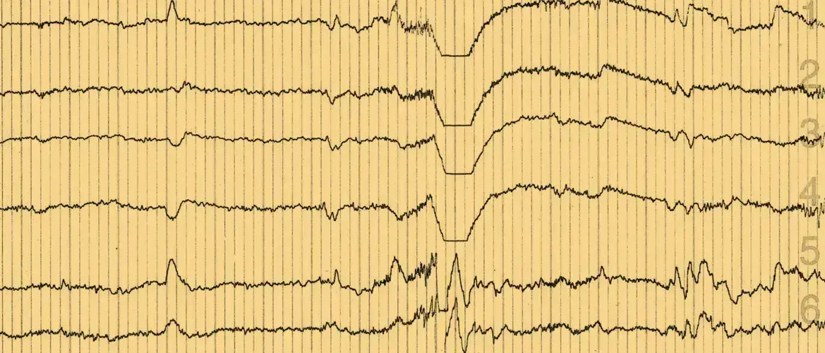
Non-convulsive epileptic activity is common in AD and is associated with the onset of cognitive decline.
비경련성 뇌전증 활성화(Non-convulsive epileptic activity)는 알츠하이머병에서 흔한 증상이며 인지기능 저하의 시작(onset)과 연관되어 있습니다.
In amyloid precursor protein (APP) transgenic mice, decreased synaptic transmission is seen alongside network excitability, which is apparent as both convulsive and non-convulsive epileptic activity. In the absence of endogenous tau, EEGs appear normal, suggesting that amyloid-beta (Aβ) requires tau to induce hypersynchrony.
아밀로이드 전구단백질(APP) 유전자 변형 생쥐에서 시냅스 전달 감소와 함께 신경망 흥분이 관찰됐는데, 이는 경련성/비경련성 뇌전증 활성화 모두에서 명확하게 관찰되는 현상입니다. 내인성 타우가 존재하지 않을 때, 뇌파(EEG)는 정상으로 나타나며 이는 베타-아밀로이드(β-amyloid)가 과동기화(hypersynchrony)를 유도하기 위해 타우를 필요로 한다는 것을 의미합니다.
APP CTF (C-terminal fragments) are elevated in patients with autosomal dominant AD; and many patients with familial AD mutations experience clinically observed seizures. 1, 2 In patients with mild cognitive impairment (MCI) or sporadic AD and epilepsy, a high proportion of seizures (>50%) are non-convulsive and seizure onset often clusters around the beginning of cognitive decline.3 Subclinical epileptic activity is detected in more than 40% of patients with early-onset, sporadic AD and appears to be associated with more rapid cognitive decline.3, 4 Other studies have also shown that measures of network dysfunction are useful biomarkers for AD.5-7
상염색체 우성 알츠하이머병 환자의 경우 아밀로이드 전구체단백질(APP) CTF (C-말단 조각: C-terminal fragments)이 증가했으며, 가족성 알츠하이머병 변이 환자 다수에서 임상적인 발작이 관찰됩니다.1, 2 경도인지 장애(MCI), 산발형 알츠하이머병, 그리고 뇌전증 환자의 경우, 발작의 다수(>50%)가 비경련성이며, 발작의 시작(onset)이 인지기능 저하가 시작되는 시점과 연관되는 경우가 많습니다.3 준임상적(subclinical) 뇌전증 활성화는 조발성 산발형 알츠하이머병 환자의 40% 이상에서 관찰되며, 이는 보다 급속한 인지기능 저하와 관련이 있는 것으로 보입니다.3, 4 그 외 연구들도 신경망 기능 장애를 알츠하이머병의 바이오마커로 이용할 수 있음을 보여주었습니다.5-7
The degree of epileptic activity in patients with AD seems to be greatest at night, when EEG detection is also most sensitive, with 1‑hour of sleep recording equivalent to 8‑hours of daytime recording.8, 9 Magneto-encephalography with EEG also appears to be more sensitive to subclinical epileptic activity in patients with sporadic AD than 24-hour video EEG.4
알츠하이머병 환자의 뇌전증 활성화는 야간에 더욱 빈번하게 발생하며, 이 시간은 뇌파검사(EEG) 결과 또한 매우 민감하게 관찰되어 수면 중 1시간 동안의 EEG 기록은 주간에서의 8시간 동안의 기록에 상당합니다.8, 9 산발형 알츠하이머병 환자의 준임상적 뇌전증 활성화 또한 24시간 비디오 뇌파검사(EEG)보다 자기뇌파검사법의 뇌파검사(EEG) 상으로 매우 민감하게 관찰됩니다.4
Antiepileptic drugs that bind the synaptic vesicle protein 2A (SV2A) have been shown to reverse hippocampal hyperactivation in the dentate gyrus/CA3 and improve performance on a pattern separation task in patients with amnestic MCI.10
시냅스 소포 단백질 2A(SV2A)에 결합하는 항전간제는 해마의 치상회(dentate gyrus)/CA3 영역에서 과활성(hyperactivation)을 역전시키는 한편, 기억력이 저하된 경도인지 장애(MCI) 환자의 패턴 분리 시험 (pattern separation task)에서 수행능력을 개선하는 것으로 확인된 바 있습니다.10
SV2A-binding antiepileptic drugs can block the network dysfunction cascade.
시냅스 소포 단백질 2A(SV2A)-결합성 항전간제는 신경망 기능장애 반응이 진행되는 것을 막을 수 있습니다.
In people with epilepsy, fast gamma oscillations are associated with the suppression of epileptic activity. Mouse and human studies of AD suggest that APP/Aβ impair fast gamma oscillations by depleting interneuron NaV1.1 sodium channels, leading to hypersynchrony and decreased cognitive function. 11, 12 In contrast, restoring NaV1.1 activity in mice has shown benefits on brain dysrhythmias and cognitive deficits, while increasing gamma oscillations led to prevention of plaque load. 11, 12
빠른 감마 진동은 뇌전증 환자의 증상 억제와 연관이 있습니다. 인간 및 생쥐를 대상으로 한 알츠하이머병 연구 결과, 아밀로이드 전구체단백질(APP)/ 베타-아밀로이드(β-amyloid)가 사이신경세포(interneuron)의 NaV1.1 나트륨채널을 감소시켜 감마 진동 속도를 낮추는 것이 확인되었습니다. 이로 인해 과동기화(hypersynchrony)가 야기되고 인지기능의 저하가 발생합니다.11, 12 반면, 생쥐의 NaV1.1 활성을 회복시키자 뇌파 율동 이상(dysrhythmias) 및 인지기능 저하가 개선되고, 감마 진동 또한 증가하여 플라크 부하(plaque load)를 예방하는 효과가 있는 것으로 확인됐습니다.11, 12
APP/ Aβ, ApoE4 and tau play a role in synaptic and network dysfunction
아밀로이드 전구체단백질(APP)/ 베타-아밀로이드(β-amyloid), ApoE4, 타우는 시냅스 및 신경망 기능장애에 영향을 끼칩니다.
ApoE4 and Tau may also play a role in hypersynchrony and subsequent cognitive impairment:
또한, ApoE4 와 타우는 다음과 같이 과동기화(hypersynchrony) 및 그에 따른 인지기능 장애에도 관여합니다.
- ApoE4 through a disruption of slow gamma activity during short-wave ripples in the hippocampus, which can be prevented if ApoE4 is eliminated. 13
해마 내 단파 방출 시 저속 감마 활동을 저해하는 ApoE4의 역할. ApoE4 제거 시 이러한 현상을 막을 수 있음.13
- Tau via a toxic gain of function or permissive overactivation induced by Aβ.14 Tau reduction also blocks the network function cascade.14, 15
독성 기능획득(toxic gain of function) 또는 베타-아밀로이드(Aβ)에 의해 유발되는 과도한 활성화(permissive overactivation)를 통한 타우의 역할. 14 타우 농도가 감소하면 신경망 기능 연쇄반응 또한 차단됨.
However a number of questions remain with regard to the best therapeutic approach. We do not know which is the most pathogenic form of tau, nor whether spreading of tau impairs critical neuronal functions or survival. Reductions in the power of delta/theta oscillations are seen in tau-ablated mice – and increases in delta/theta power predict progression to AD in people with subjective impairment.16, 17
그러나 최적의 치료적 접근을 위해서는 아직 풀어야 할 과제가 많습니다. 어떤 형태가 가장 병원성이 높은 타우의 형태인지, 타우의 확산이 주요 신경 세포의 기능 또는 생존에 장애를 일으키는지 알지 못합니다. 타우를 제거한 쥐는 델타/세타 진동 강도가 감소하는 것이 관찰되었는데, 델타/세타 강도 증가는 주관적으로 인지능력 저하를 자각하고 있는 사람이 알츠하이머병으로 진행되는 정도를 예측할 수 있도록 해주는 예측인자입니다.16, 17
본 자료는 Global Lundbeck 의학부에서 선별한 학술대회 콘텐츠이며, 한국룬드벡의 의견과 다를 수 있습니다